INTRODUCTION
The presence of shoulder pain disrupts shoulder mobility, causing stiffness and weakness that lead to a marked decrease in a person’s ability to work, practice sports, and even perform everyday activities.
There are many different conditions that can cause shoulder pain, and they must be differentiated before starting any treatment.
Conditions that cause shoulder pain
Conditions specific to the shoulder region
-
Subacromial impingement, which causes:
- Subacromial bursitis, or subdeltoid bursitis.
- Tendon injuries: acute supraspinatus tendinitis, supraspinatus tendinosis, calcific or non-calcific, or tendinosis of the proximal biceps tendon.
- Rotator cuff tear, partial or complete.
- Shoulder instability. This is generally related to a previous glenohumeral dislocation.
- Adhesive capsulitis. This is seen after prolonged immobilization or in patients with diabetes and is characterized by pain and marked stiffness.
- Glenohumeral or acromioclavicular osteoarthritis. These are degenerative conditions related to age and are typical of patients older than 50 years.
Pathology in related organs
- Cervical pathology with referred pain, most commonly due to a degenerative process.
- Tumors of different types.
The most common shoulder pain in the young athlete
The most common shoulder pain in the young athlete is caused by supraspinatus tendinitis. Its acute form is typical of the young, vigorous patient who performs load-bearing physical activities with excessive use of the shoulder, causing increased friction, either at work or in sports. This injury is related to “subacromial impingement,” a very common condition that causes tendon injuries and bursal injuries in the subacromial space.
For this reason, we will focus on this topic and briefly discuss other conditions that must be ruled out.
Pathology of the subacromial space
In the medical literature, multiple terms have been used to describe pathology of the subacromial space of the shoulder, mainly because it involves different elements within the space, either alone or in combination, with a clinical picture produced by mixed injuries. Thus, the following terms may be found:
- Subacromial impingement syndrome
- Subacromial pain syndrome
- Subacromial syndrome
- Rotator cuff syndrome
- Supraspinatus tendinopathy or tendinitis
- Partial or complete rotator cuff tear
- Calcific tendinitis
- Subacromial bursitis
To standardize terminology, the general term “rotator cuff disease” has been proposed to refer to disorders of the elements of the subacromial space, regardless of their cause and specific anatomical location; however, there is still no consensus on this. (1)
Therefore, in this article we will use the most common name, “subacromial impingement.”
Subacromial Impingement in the Athlete
Concept
Subacromial impingement (SAI) is defined as increased pressure exerted on the elements inside the subacromial space (SAS); these elements become trapped between the acromion on one side and the humeral head on the other, initially undergoing acute inflammatory changes that later become chronic and degenerative.
Subacromial impingement is the most common pathology that causes shoulder pain in young athletes, especially those who perform throwing, repetitive movements, or weightlifting above the head. In this age group, impingement is considered to be caused by overuse, and its acute form is the most common; it has been suggested that young athletes may have lax shoulders or scapular dyskinesia as an underlying disorder associated with impingement. For this reason, it should be considered a separate entity from degenerative shoulder pathologies.
In the general population, it is the cause of more than 50% of shoulder pain cases, and many different occupations are associated with it, especially those requiring heavy lifting, repetitive movements in awkward positions, or exposure to vibration.
The incidence of subacromial pain related to impingement increases with age, with chronic degenerative forms predominating and peak incidence occurring in the sixth decade of life. The pathology is associated with shoulder muscle weakness, altering activities of daily living.
Basic Anatomy of the Subacromial Space
The SAS is bounded inferiorly by the humeral head and superiorly by the elements that form the so-called “coracoacromial arch,” which are: the anterior border and inferior surface of the anterior third of the acromion, the coracoacromial ligament, the acromioclavicular joint, and the coracoid process. The height of the space varies from 1.0 to 1.5 cm and physiologically decreases with abduction, flexion, and internal rotation of the shoulder.
The SAS contains the rotator cuff tendons, from front to back: subscapularis, supraspinatus, infraspinatus, and teres minor, and the tendon of the long head of the biceps, as well as the serous sac or subacromial bursa that separates them from the inferior border of the acromion. Because of its position in the SAS, the supraspinatus tendon is the one most often affected in cases of impingement.


a) Drawing. b) Neer and Poppen Y projection.
Red oval: subacromial space. Blue oval: glenoid cavity. Black circle: humeral head. Black line: acromial arch.
A- Acromion, AC- Acromioclavicular joint, LCA- Coracoacromial ligament, TB- Proximal biceps tendon, C- Coracoid process, E- Scapula, H- Humerus.

- 1- Coracoacromial ligament
- 2- Subacromial space
- 3- Joint capsule through the rotator interval
- 4- Acromion
- 5- Infraspinatus
- 6- Supraspinatus
- 7- Subscapularis
- 8- Humerus
- 9- Proximal biceps tendon
- 10- Clavicle
- 11- Coracoid
https://doi.org/10.1016/S1286-935X(08)70919-5
ETIOLOGY OF IMPINGEMENT (3,7,8)
Impingement of the subacromial space may be divided into external or internal:
- External impingement, in which the alterations are outside the space. Impingement is caused by elements that form the acromial arch, which mechanically compress the soft tissues located within the SAS.
- Osteophytes, or bone spurs, on the acromion or changes in its morphology, such as a hooked acromion.
- Degenerative changes of the acromioclavicular joint.
- Degenerative changes of the coracoacromial ligament.
- Internal impingement, caused by rotator cuff damage. Impingement occurs when the rotator cuff tendons invade the humeral head; the articular side of the cuff, posterolateral portion, is pinched against the glenoid rim and the labrum, in its posterosuperior portion, when the shoulder is in abduction and external rotation, in an overhead throwing position.
The consensus is that in young patients, acute pathology of the supraspinatus and, in general, of the rotator cuff may be due to intense trauma, falls, or glenohumeral dislocation, or may be related to repetitive and forceful activities in sports or work; the existence of a genetic muscular predisposition is suspected. Repeated injury or tension causes swelling and pain; once the muscle is inflamed, pain appears or worsens when the arm is lifted in abduction because impingement occurs over the cuff.
In people older than 40 years, age-related vascular changes increase the normal hypovascularity of the cuff and cause a chronic degenerative tendon process; in this phase, cuff rupture prevents containment of the humeral head and allows its migration and elevation, which decreases the subacromial space. This feature is observed more frequently in people older than 60 years.
Shoulder impingement is also classified according to its cause, as primary or secondary.
- Primary, when there is structural narrowing, such as acromial deformity.
- Secondary, when impingement begins with movement due to muscle weakness, of the cuff or of the trapezius and serratus anterior.
Of the alterations external to the space that have been related to subacromial pathology, much interest has been placed on changes in acromial morphology. According to Bigliani, there are three types of acromion: Type I, flat surface. Type II, curved surface. Type III, hook-shaped. Neer’s concept regarding the relationship between acromial shape and the presence of subacromial impingement is classic, with a higher frequency observed in the presence of hook deformity. However, it is not clear whether this is the cause or the effect of the degenerative rotator cuff process. (figure 4) (3)
Likewise, controversy persists as to whether damage to the rotator cuff tendons leads to impingement, intrinsic mechanism, or whether impingement causes tendon damage, extrinsic mechanism. The intrinsic theory has been gaining ground.

PATHOLOGY
When the SAS is impinged, an inflammatory process begins in the subacromial bursa and the rotator cuff tendons, mainly the supraspinatus and the biceps, acute tendinitis and acute subacromial bursitis. As impingement becomes constant, acute tendinitis becomes chronic, leading to tendon degeneration, or tendinosis, partial or complete rotator cuff tear, and finally, a subacromial abrasion syndrome produced by continuous rubbing of the humeral head against the acromion. This process begins at the supraspinatus because it is the most vulnerable due to its anatomical position. (figure 5)
Neer’s classification is based on these concepts. (3)
Neer Classification
Lesions of the supraspinatus tendon are divided into 3 stages according to their evolution:
- Stage 1: Reversible tendon edema and hemorrhage due to overuse; younger than 25 years.
- Stage 2: Tendinitis and fibrosis with recurrent pain during activity; patients between 25 and 40 years.
- Stage 3: Bone spurs and tendon tear with progressive disability; in patients older than 40 years.


It has been suggested that the poor vascularization of the insertion of the supraspinatus tendon may be an important factor in the pathogenesis of degenerative rotator cuff tears, beginning in the so-called “critical zone.” The source of pain is the free nerve endings found in the bursa, and its intensity is related to the degree of damage to the supraspinatus tendon.
Because of its anatomy, the terms rotator cuff tendinitis and rotator cuff tear are used as synonyms for supraspinatus tendinitis and supraspinatus tear, and they are used interchangeably. Nevertheless, in general, tendinitis involves the supraspinatus, while rupture involves the rotator cuff.
We can conclude that, even when the initial acute injury involves only one element, later all those found within the subacromial space become compromised, which is why these pathologies become clinically confused, making differential diagnosis difficult.
These pathologies are:
- Inflammation of the subacromial serous sac, or “bursitis.”
- Supraspinatus tendinopathy, tendinitis or tendinosis.
- Rotator cuff tear of the shoulder.
- Tendinopathy of the long head of the biceps, tendinitis or tendinosis.
Subacromial bursitis
The term indicates inflammation of the subacromial sac or bursa and may be due to impingement. It is the first step in compressive pathology because the bursa is a barrier that separates the tendons within the SAS from the other elements that limit it above, the acromial arch.
Supraspinatus tendinopathy
Tendinopathy comprises two phases: the initial acute inflammatory phase, or tendinitis, and the chronic degenerative phase, or tendinosis; acute lesions have a greater probability of complete regeneration.
Tendinopathy may be calcific or non-calcific. Calcific tendinopathy presents with calcium deposits inside the rotator cuff tendons and adjacent bursa, visible on X-rays, and may produce symptoms of a rotator cuff tear.
In tendinopathy, poor healing of the extracellular matrix is observed microscopically, with collagen degeneration and increased levels of proteoglycans and type III collagen; these fibers lose their hierarchical structure and become more irregular and spaced out.
Tendinosis involves changes in the structure and arrangement of collagen fibers and neovascularization within the tendon. (9)
The neovascularization process is typical of tendon injuries and is crucial in the tendon healing process, with the presence of growth factors and the emergence of stem cells that form tenocytes; however, it plays a dual role in tendon disorders because it is also an important component of tendon tissue degeneration when regeneration and healing are incomplete.
In chronic degenerative tendinopathy, a neovascularization process exists and, although theoretically the adjacent growth of nerve endings is responsible for pain, there is not always a connection between neovascularization and pain, and it has been observed in tendons of asymptomatic athletes.
Rotator cuff tear
Pathology
When impingement becomes permanent, it eventually causes rupture of the tendons that form the rotator cuff, beginning with the supraspinatus, since, due to its position beneath the acromion, it is the most vulnerable and begins in its least vascularized portion, called the “critical zone,” in its deep part, which is in contact with the humeral head, until it tears through its entire thickness and communicates the articular surface with the bursal surface.
The inflammatory or degenerative lesion of the supraspinatus is confused with rotator cuff tear, because both produce pain on palpation and on shoulder movement in abduction.
When the cuff tears, the humeral head ascends and rests against the acromion and the coracoacromial ligament, increasing pressure and friction against the acromial arch, causing abrasion that is initially asymptomatic and later causes pain with shoulder movement.
Classification (figure 6)
- Partial tear, “non-transfixing.” The cuff tear begins in the deep fibers and causes shoulder pain and disability.
- Full-thickness tear, transfixing. As the cuff tear increases, pain decreases, but difficulty raising the arm above the head progressively increases, until it becomes impossible when the cuff tears completely.

Proximal biceps tendinopathy
The most frequent cause of proximal biceps tendinitis is subacromial impingement; another way it may occur is friction within the bicipital groove due to continuous rubbing of the tendon. It progresses to tearing and may reach rupture. This tendinopathy commonly accompanies other lesions within the subacromial space, shoulder instability, and injury at its origin, in the anterosuperior supraglenoid region of the humerus, known as a SLAP lesion. (figure 7)

Risk factors
Painful shoulder caused by subacromial pathology is common in sports that involve moving the arms above the head, whether through throwing, lifting heavy loads, or repetitive movements, such as tennis, basketball, baseball, weightlifting, and swimming, in which intensive training may cause pain. The same occurs in people who work in similar circumstances, such as painters, electricians, bricklayers, and warehouse workers.
Weakness of the muscles, ligaments, and tendons of the shoulder is considered another risk factor, since the shoulder is a very mobile joint complex with little stability, which depends on muscular action. (figure 8)


CLINICAL PICTURE (3,8,10)
Impingement may cause combined injuries in one or more elements within the SAS, such as bursitis, supraspinatus tendinitis, biceps tendinitis, or rotator cuff tendinosis. For this reason, clinical findings overlap and it is often impossible to reach a definitive diagnosis based only on clinical findings.
The acute phase generally occurs in a young patient, younger than 40 years, with a history of using the shoulder in activities, whether sports or work, above the head, with intense pain of sudden onset in the lateral part of the shoulder after these activities.
A chronic pain picture more frequently affects patients older than 40 years, and in these cases, pain is related to a degenerative process.
Symptoms
The predominant complaints are pain and disorders of shoulder function.
Pain. In the initial acute phase, pain is intense and is referred by the patient to the lateral area of the shoulder, below the acromion. The main characteristic of the pain is that it increases when the arm is lifted away from the trunk, in abduction, and brought above the head, especially with load; pain may also occur when lying on the affected side, more commonly at night, causing sleep disturbances.
In the chronic stage, pain begins gradually and insidiously, over weeks or months, often radiating to the proximal part of the arm along its lateral border; it is accompanied by shoulder weakness and decreased mobility.
When there is clear weakness without pain, it may indicate a complete rotator cuff tear; however, this finding is not always present.
If the proximal biceps tendon is involved, pain also includes the anterior shoulder region and may radiate toward the anterior part of the arm.
In SLAP lesions, which affect the origin of the proximal biceps tendon, onset may be acute, with pain in the anterior part of the shoulder, accompanied by clicking, locking, or catching during shoulder movements. However, onset may also be insidious, predominating at night and radiating to the anterior part of the arm, accompanied by a sensation of weakness in the shoulder.
The inflammatory process may involve the subscapularis muscle, which should be suspected if the pain is anterior, adjacent to the proximal biceps tendon. This can be confirmed through the appropriate clinical tests.
Finally, when the patient refers pain to the posterior part of the shoulder, degenerative pathology should be considered and referred pain from the cervical spine should be ruled out.
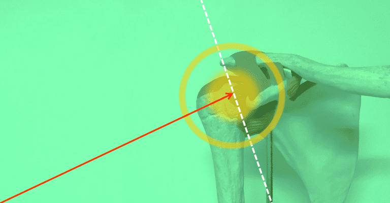
Functional disorders. Pain leads to muscle weakness with decreased mobility and strength of the affected shoulder when bringing it above the head, especially when lifting a load, which limits shoulder function. Pain typically appears between 60° and 120° of shoulder abduction, the “painful arc,” and disappears with greater elevation. (Figure 9)
Adhesive capsulitis causes limitation of shoulder rotations and may be very painful in its acute phase. It is more common in patients older than 60 years with diabetes mellitus and often affects both shoulders at the same time or separately.

Signs
The patient’s physical examination must be thorough, trying to define the injured elements, although this tends to be difficult and sometimes impossible.
All elements that make up the shoulder joint are palpated to identify painful points. In SAI, pain is elicited by digital pressure on the lateral side of the shoulder, over and below the acromial process, the site of the SAS. If proximal biceps tendinopathy is present, pain appears when pressing on the anterior part of the shoulder, in the bicipital groove. (figure 10)
Observing scapular mobility is important; normally, the scapula begins to move during abduction until the arm abducts to 90 degrees; when scapular dyskinesia exists, its movement begins much earlier. It may appear when shoulder pain has become chronic.


Basic clinical maneuvers (3, 8, 10)
Tests for the supraspinatus
Jobe test, empty can test. This test looks for the appearance of pain and weakness during shoulder abduction against gravity in internal rotation, in the scapular plane: the arm is raised in 30° antepulsion-abduction and internal rotation, placing it in the plane of the scapula with the thumb pointing toward the floor. Pain appears or worsens between 60° and 120° of abduction, when the supraspinatus becomes trapped under the acromion, and disappears with greater abduction. Next, the examiner tries to lower the arm against the patient’s resistance, causing the pain to appear again. (figure 11)
Drop arm test. The patient is placed with the shoulder at 90 degrees of abduction and internal rotation in the scapular plane. The examiner supports the limb and then instructs the patient to lower the arm slowly toward the side of the body. A positive test includes the patient’s inability to maintain the abducted shoulder position and/or inability to lower the arm in a controlled manner. A positive result raises suspicion of rotator cuff tear. (figure 11)
Test for the infraspinatus
Patte test. With the arm against the body and the elbow flexed at 90º, the patient is instructed to perform external rotation against the examiner’s resistance, causing shoulder pain.
Test for the teres minor
Hornblower test. The shoulder is placed in 90° abduction and external rotation with support, with the elbow flexed at 90°. It is positive if the patient cannot maintain this position when support is removed and the arm moves into internal rotation; complete rotator cuff tear is suspected.


Tests for the subscapularis (figure 12)
Hawkins test. With the shoulder in antepulsion, resistance is applied against internal rotation, provoking pain. If pain occurs when moving into external rotation, the infraspinatus is examined.
Belly press test. The standing patient places the elbow at 90 degrees of flexion and presses the palm against the abdomen while the examiner brings the patient’s elbow in front of the plane of the trunk, forward. The test is positive if the patient cannot resist the examiner’s force, due to weakness or the appearance of pain. Another way to assess this is to ask the patient to maintain the elbow in this position when the examiner removes the supporting force; inability to do so suggests subscapularis tear.
Gerber test, lift-off test. There is pain and decreased strength during the final degrees of internal rotation with the elbow flexed at 90° and the hand behind the back.


Tests for proximal biceps tendinopathy and SLAP lesions (figure 13)
De Anquin test. Shoulder rotations provoke pain in the bicipital groove.
Speed test. The shoulder is flexed against resistance, with the elbow extended and the forearm in supination, causing pain in the bicipital groove. It is the most reliable sign of bicipital tendinitis.
Uppercut test. The affected shoulder is placed in neutral position, the elbow is flexed at 90 degrees, the forearm is supinated, and the patient makes a fist. The examiner instructs the patient to perform an uppercut punch, bringing the arm forward and upward, while placing a hand over the patient’s fist to resist the movement. It is positive if pain or a painful click appears over the anterior part of the shoulder, next to the bicipital groove.
Yergason test. With the elbow flexed at 90 degrees and the forearm in pronation, the patient supinates the elbow and abducts the shoulder while the examiner manually resists. It is positive if it elicits pain over the bicipital groove and/or subluxation of the tendon.


Neer test to rule out rotator cuff tear. This consists of injecting a local anesthetic into the subacromial space to eliminate pain. If, when pain decreases, the patient can raise the shoulder in abduction above the head, this indicates that the rotator cuff is not torn and that it is only an inflammatory process.
Imaging studies (3,5)
Standard shoulder X-rays include 2 views: AP and Y projection. (figures 14 and 15)
On the AP shoulder X-ray, the following are determined: (figure 14)
Radiographic measurements and their relationship with shoulder pathologies
- The critical shoulder angle of Moor (CSA) is the angle formed by a line connecting the superior and inferior borders of the glenoid and another line going from the inferior border of that glenoid to the inferior and lateral border of the acromion. The normal CSA ranges from 30° to 35°.
- The acromial index of Nyffeler (AI) is the relationship between the distance from the glenoid to the lateral border of the acromion (GA) and the distance from the glenoid to the lateral border of the humeral head (GH). AI = GA/GH. The normal AI is 0.74.
Both the CSA and the AI are measured in the coronal plane and describe the lateral displacement of the acromion in relation to the humeral head. In theory, a large lateral acromial extension, with a CSA greater than 35° and a high AI greater than 0.74, may cause excessive shear forces from the deltoid on the glenohumeral joint and increase the load on the rotator cuff tendons, increasing the risk of tears. Conversely, a CSA below 30° and an AI lower than 0.74 indicate a shorter acromion with less coverage of the humeral head; this situation would result in increased compressive forces in the glenohumeral joint with increased risk of osteoarthritis.
An important source of error is the radiographic technique. Accurate measurement requires an exact frontal X-ray because a small deviation in the radiograph affects the CSA, since its precision depends on the relationship between the scapula and the X-ray beam, which varies with the patient’s position and posture.
A close relationship has been reported between supraspinatus and rotator cuff tendinopathy and the CSA and AI, with an increase in frequency in shoulders with a CSA greater than 35°, since as the acromion becomes more lateral, friction with the rotator cuff increases; the larger the CSA, the larger the rotator cuff tear. Regarding the acromial index, the closer it is to one, the greater the coverage of the head and the greater the risk of tendinopathy. Nevertheless, controversy persists, with reports reaching conclusions both for and against this relationship. (11,12)
X-rays are also useful for detecting calcific tendinitis and ruling out other associated pathologies, such as osteoarthritis; the scapular Y view allows evaluation of the humeral head in the glenoid cavity and the shape of the acromial process. (figure 15)
The ideal imaging study for detecting tendon injuries is magnetic resonance imaging; however, due to its high cost, ultrasonography is used more frequently.
There is not always a direct relationship between shoulder pathologies detected by imaging and the patient’s clinical findings; imaging studies may detect pathologies in asymptomatic individuals, but may also show lesions that can correlate with pain, more frequently in cases of rotator cuff tear and adhesive capsulitis.
Studies have shown that numerous pathologies may coexist in the same individual.


BIBLIOGRAPHY
- Page MJ, Green S, McBain B, Surace SJ, Deitch J, Lyttle N, Mrocki MA, Buchbinder R. Manual therapy and exercise for rotator cuff disease. Cochrane Database Syst Rev. 2016 Jun 10;2016(6):CD012224. doi: 10.1002/14651858.CD012224. PMID: 27283590
- Consigliere P, Haddo O, Levy O, Sforza G. Subacromial impingement syndrome: management challenges. Orthop Res Rev. 2018 Oct 23; 10:83-91. doi: 10.2147/ORR.S157864. PMID: 30774463.
- Creech JA, Silver S. Shoulder Impingement Syndrome. [Updated 2022 Apr 21]. In: StatPearls [Internet]. Treasure Island (FL): StatPearls Publishing; 2022 Jan-.
- Allen GM. The diagnosis and management of shoulder pain. J Ultrason. 2018;18(74):234-239. doi: 10.15557/JoU.2018.0034. PMID: 30451406; PMCID: PMC6442215.
- Garving C, Jakob S, Bauer I, Nadjar R, Brunner UH. Impingement Syndrome of the Shoulder. Dtsch Arztebl Int. 2017 Nov 10;114(45):765-776. doi: 10.3238/arztebl.2017.0765. PMID: 29202926; PMCID: PMC5729225.
- org [Internet]. Cologne, Germany: Institute for Quality and Efficiency in Health Care (IQWiG); 2006-. Shoulder pain: Overview. 2020 Feb 13.
- Tapscott DC, Varacallo M. Supraspinatus Tendonitis. [Updated 2022 Feb 12]. In: StatPearls [Internet]. Treasure Island (FL): StatPearls Publishing; 2022 Jan-.
- Varacallo M, El Bitar Y, Mair SD. Comprehensive Shoulder Evaluation Strategies. [Updated 2022 May 1]. In: StatPearls [Internet]. Treasure Island (FL): StatPearls Publishing; 2022 Jan-.
- Jaworski Ł, Zabrzyńska M, Klimaszewska-Wiśniewska A, Zielińska W, Grzanka D, Gagat M. Advances in Microscopic Studies of Tendinopathy: Literature Review and Current Trends, with Special Reference to Neovascularization Process. J Clin Med. 2022 Mar 13;11(6):1572. doi: 10.3390/jcm11061572. PMID: 35329898; PMCID: PMC8949578.
- May T, Garmel GM. Rotator Cuff Injury. [Updated 2022 May 1]. In: StatPearls [Internet]. Treasure Island (FL): StatPearls Publishing; 2022 Jan-.
- Björnsson Hallgren HC, Adolfsson L. Neither critical shoulder angle nor acromion index were related with specific pathology 20 years later! Knee Surg Sports Traumatol Arthrosc. 2021 Aug;29(8):2648-2655. doi: 10.1007/s00167-021-06602-y. PMID: 34009456; PMCID: PMC8298350.
- Hsu TH, Lin CL, Wu CW, Chen YW, Vitoonpong T, Lin LC, Huang SW. Accuracy of Critical Shoulder Angle and Acromial Index for Predicting Supraspinatus Tendinopathy. Diagnostics (Basel). 2022 Jan 22;12(2):283. doi: 10.3390/diagnostics12020283. PMID: 35204373; PMCID: PMC8871498.
Discover more from Dr. Roberto García
Subscribe to get the latest posts sent to your email.