Orange background with the Spanish word “ESGUINCE” (sprain) in white, the letter “N” highlighted in yellow, and decorative lines and a circle.

General Information About Sprains

A sprain is a ligament injury caused by stretching, with partial or complete rupture. This article explains joint stability, sprain grades, causes, clinical signs, imaging studies, RICE treatment, cryotherapy, kinesiotaping, and physical therapy.
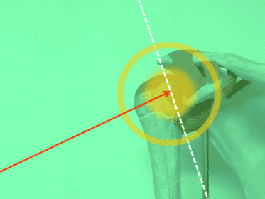
Anatomical image of a human shoulder on a green background, with a red arrow pointing to the glenohumeral joint and white and yellow reference lines.

Subacromial Impingement in Athletes

Subacromial impingement is one of the most common causes of shoulder pain in young athletes. This article explains its anatomy, causes, symptoms, clinical tests, imaging studies, and related rotator cuff conditions.

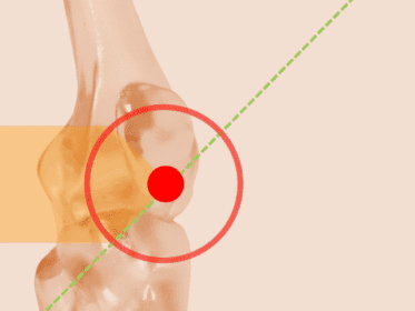
Anatomical image of the human elbow on a beige background, with a red circle highlighting the joint and a green diagonal line crossing the marked area.

Understanding Jumper’s Knee: Causes, Treatment, and Recovery from a Specialist’s View

Jumper’s knee is a common yet complex condition affecting athletes of all levels. This in-depth guide by Dr. Roberto García García explains the anatomy, causes, symptoms, and effective treatment options for patellar tendinopathy. Grounded in medical expertise and supported by research, the post offers clarity, solutions, and hope for long-term recovery.

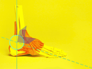
Anatomical model of the foot and ankle, with muscles highlighted in red and purple on a bright yellow background and blue lines crossing the joint, which is marked with a blue circle.

Ankle sprain: grades, treatment and when to see a doctor

Ankle sprains are one of the most common orthopedic injuries. In this comprehensive guide, Dr. Roberto García García explains how to diagnose, classify, and treat ankle sprains—ranging from mild to severe. Backed by medical research and decades of clinical experience, this post is essential for patients, athletes, and professionals alike.

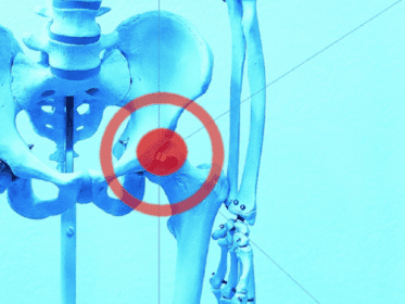
Bone model of the pelvis and femurs, with the right hip joint highlighted by a red circle on a blue background.

Practical Guide to Identifying Hip Dislocations on X-Rays

Dr. Roberto García García presents a clear method for identifying hip dislocations on X-rays using quadrant-based lines. This clinical guide, adapted from his textbook, includes real cases and radiographic images. Designed to support medical students and professionals in mastering trauma-related hip injuries and diagnostic interpretation.