Roberto García García – Orthopedic and Trauma Specialist
February 2022
Ankle sprains are very common injuries that can occur when the foot twists while walking, running, jumping, or during sports activities. The severity of the injury depends on the load the ankle bears and the speed at which the twisting motion occurs. For instance, a sprain resulting from jumping from a considerable height tends to be more severe than one caused by a misstep while walking. The symptoms, physical findings during examination, and required treatment all depend on the degree of injury.
An ankle sprain is defined as “the stretching or partial to complete tearing of one or more ligaments of the ankle joint, caused by an involuntary twisting motion that exceeds the joint’s normal range of movement.”
Since lateral sprains are the most common type, this article will focus on that specific presentation.
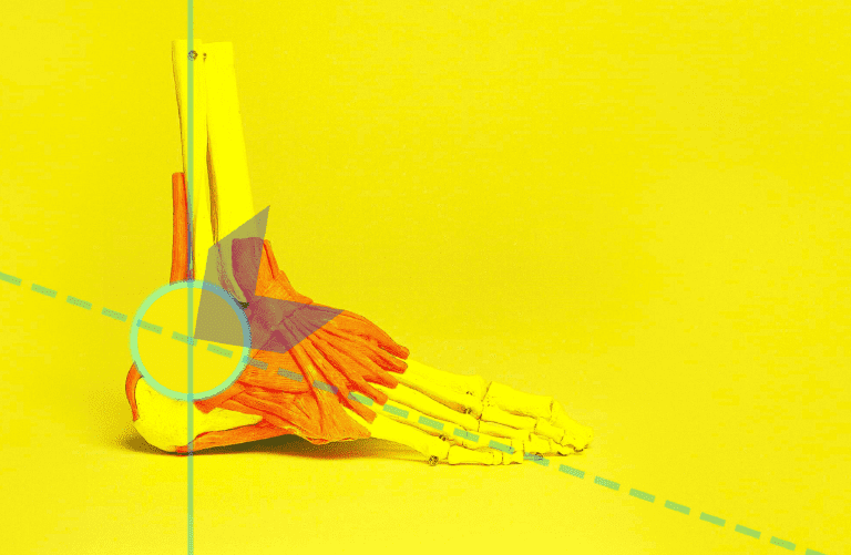
Some Ankle Anatomy (1)
To understand this topic, it’s important to review a few basic anatomical concepts of the ankle.
The ankle region includes the distal end of the tibia and fibula (also called the peroneus), and their joint surface with the talus, forming the tibiofibular-talar joint.
However, in the fields of Traumatology and Orthopedics, the ankle region is divided into two areas for the purpose of studying injuries: the tibial plafond, which is the distal meta-epiphysis of the tibia, and the malleolar region, which includes the medial (tibial) and lateral (fibular) malleoli.
Figure 1. Ankle anatomy. A) Malleolar region and its relationship with the talus. B) Frontal view of the tibiofibular-talar joint.
This surgical division is based on the differences in fracture types, mechanisms of injury, and the corresponding treatment methods required.

Figure 1. Frontal (AP) X-ray of the ankle region. Injuries specific to the ankle typically occur in the malleolar zone. MP – lateral or fibular malleolus, MT – medial or tibial malleolus.
Ankle stability is provided by three ligament systems:
- The lateral ligament complex. In the malleolar region, these ligaments connect the tibia (medially) and fibula (laterally) to the hindfoot bones (talus and calcaneus).
- The syndesmotic ligaments. Located in the tibial plafond, these ligaments join the distal tibia and fibula, forming the tibiofibular syndesmosis.
- The medial deltoid ligament, both superficial and deep layers.
The lateral ligaments are made up of three bands from front to back: anterior talofibular ligament (ATFL), calcaneofibular ligament (CFL), and posterior talofibular ligament (PTFL).
The syndesmotic ligaments primarily include the anterior and posterior tibiofibular ligaments.


Figure 2. A) AP X-ray showing the three ligament bundles of the ankle: medial, lateral, and syndesmotic. B) Lateral anatomical drawing. PTFL – posterior talofibular ligament, ATFL – anterior talofibular ligament, CFL – calcaneofibular ligament.
- Sprains
- Fractures
- Sprains most commonly affect the lateral ligaments, followed by the medial and syndesmotic ligaments.
- A significant number of fractures result from the same injury mechanism as a lateral sprain but involve a higher-energy force.
Lateral Sprain
A lateral sprain results from a forced and sudden inversion of the ankle (twisting inward) while walking or during sports activities. Most commonly, the ankle also undergoes plantarflexion (with more pressure on the forefoot than the heel), leading to injury of the ATFL, which inserts in the anterior region of the ankle. If the ankle is in dorsiflexion (with more pressure on the heel, which is less common), the CFL may be injured due to its insertion in the lower medial part of the ankle. The PTFL is very strong and located in the posterior ankle, and is rarely injured.
The severity of the sprain depends on both the force placed on the ankle and the speed of the motion. A mild load during walking may cause a first-degree sprain, while greater force (e.g., jumping or running) may lead to a more severe, second- or third-degree sprain. A high-energy mechanism can cause an inversion fracture. (Figure 3)


Figure 3. A) Ankle inversion with slight plantarflexion. B) In a third-degree sprain, both the ATFL and CFL are torn.
Classification
The typical classification of a sprain generally depends on the severity of the ligament injury and is divided into degrees (stretch, partial tear, and complete tear). In the case of a sprain involving the lateral collateral ligament of the ankle, which consists of three separate bundles, the classification also refers to the number of affected fascicles (known as the Sangeorzan classification).
- TYPE I: Ligament strain affecting all three bundles, without joint laxity.
- TYPE II: Complete tear of the ATFL and partial tear of the CFL, with moderate laxity. PTFL remains intact.
- TYPE III: Complete rupture of the ATFL, CFL, and the joint capsule, leading to clear laxity and instability.
Clinical Presentation
Symptoms
Symptoms and signs typically appear on the lateral and anterior sides of the ankle. The most common symptom is pain, which can vary in intensity and is often sharp, worsening with movement. Variable swelling may be present, and bruising or discoloration may appear quickly. In severe cases, there may be an inability to walk or move the ankle.
The clinical signs depend on the severity and type of sprain:
- In a first-degree sprain, pain and swelling are mild, and the patient is able to walk. There are no signs of instability or functional impairment.
- In a second-degree sprain, pain is more intense and worsens with weight-bearing, causing the patient to avoid putting weight on the affected limb while walking. Moderate swelling, bruising (ecchymosis), and functional limitation are present. However, the patient is still able to take at least four steps.
- In a third-degree sprain, pain, swelling, bruising, deformity, and functional impairment are severe, resulting in an inability to bear weight or walk. Clinical signs of ankle instability are evident. (Figure 4)


Fig 4. Clinical presentation of a lateral ankle sprain. A) First-degree sprain with mild swelling. B) Second-degree sprain with visible submalleolar bruising.
Signs
The “eggshell sign” (also known as McKenzie’s sign) refers to a small, well-defined, ovoid area of swelling. It appears only in the early phase of moderate to severe sprains. As swelling progresses, the sign typically disappears, which explains why it is rarely observed in later stages.
When clinical signs suggest a severe sprain, it is important to assess for instability, as this may indicate a complete ligament rupture.
Tests for Lateral Ligament Injury
a) Suction or Opening Test (Watson-Jones test): This test involves forced inversion of the ankle into a varus-equinus position. A groove may appear beneath the lateral malleolus, as if the skin is being suctioned, and the joint visibly opens—indicating complete rupture of both the anterior talofibular ligament (ATFL or LPAA) and the calcaneofibular ligament (CFL or LPC), consistent with a third-degree sprain. This test can be performed while a radiograph is being taken to confirm instability. (Figure 5)


Figure 5. Suction test demonstrating ankle instability due to lateral ligament rupture. A) Clinical photo during the maneuver. B) Radiographic image.
b) Talus Tilt Test. With the ankle held in a neutral position, the talus is moved into adduction and abduction. The test is positive if inversion increases by 5° to 10° compared to the uninjured side, suggesting injury to the CFL. When performed in plantar flexion, it assesses the ATFL.
c) Anterior Drawer Test (Castaing and Deplace): With the knee and ankle at 90°, the foot is moved forward. Displacement indicates rupture of the ATFL.
d) Vega’s Sign. The anterior drawer test is considered positive only if performed in plantar flexion, suggesting micro-instability. It is used when instability is suspected but the standard anterior drawer test at 90° is negative.
How to Identify a Sprain and Differentiate It from a Fracture
(8) The symptoms reported by the patient and observed during examination in moderate or severe sprains are often similar to those of an ankle fracture. Differentiation must be made by palpating the site of pain.
- Pain from a ligament injury appears when pressing below the malleolus, where the swelling and bruising are most pronounced.
- In the case of a fracture, the pain is more intense directly over the affected malleolus. This should be examined by palpating the last six centimeters of the malleoli.
- Pain from a ligament injury tends to subside within 3 seconds of sustained pressure; pain from a fracture persists for longer than 3 seconds.
According to the Ottawa Ankle Rules, radiographic studies are required to rule out bone injury and/or instability after traumatic ankle injury, based only on two criteria:
- If the patient is unable to bear weight immediately after the injury and during the clinical examination (unable to take four steps).
- If there is significant tenderness upon palpation of the last 6 centimeters of one or both malleoli.
Treatment
Initial Treatment
Regardless of the injury grade—but especially in second- and third-degree sprains—the initial phase (inflammation phase, first 72 hours) focuses on reducing pain, swelling, and edema.
Since Mirkin’s 1978 report, the standard acute-phase treatment has been the comprehensive “RICE” protocol, an acronym for Rest, Ice, Compression, and Elevation. Later, a fifth element—Protection—was added, evolving into “PRICE.” While evidence of its effectiveness is limited, it has shown short-term benefits for pain control.
Figure 6. Example of RICE treatment applied to acute injuries.
- Rest. Keeping the injured limb at rest is essential for second- and third-degree sprains.
- Ice (Cryotherapy). Ice is useful only during the first 48 hours. It helps prevent fluid leakage and edema. After day two, it offers no additional benefit. Ice should never be applied directly to the skin—crushed ice is best, as it molds to the injured area. See the corresponding section in the general article on sprains for further guidance. The use of phase-change material cooled to 15°C, originally designed for muscle injuries, has also been adapted for sprains.
- Compression Bandage. The goal is to increase extravascular pressure and improve circulation, reducing swelling. Bandaging in a figure-eight around the ankle prevents excessive anterior pressure, which could worsen edema or cause skin necrosis. Various bandages are available to provide firm, even, and consistent compression. Padded bandages (e.g., Jones dressing) allow greater pressure but reduce cryotherapy effectiveness.
- Elevation of the Limb. Elevating the affected leg improves venous return and decreases swelling.
These measures are complemented by nonsteroidal anti-inflammatory drugs (NSAIDs) and supportive physiotherapy.
As for protection, the use of a splint or brace to support the ligaments remains controversial. A plaster cast may inhibit effective cryotherapy and compression, limiting its usefulness.

Figure 6. The “RICE” method for managing acute sprains.
Comprehensive Treatment by Sprain Grade
First-Degree Sprain
As this is a mild ligament injury, the RICE method is optional. Partial immobilization should be applied immediately, and the patient is encouraged to begin movement and weight-bearing right away, while protecting the ankle with an elastic bandage reinforced with adhesive strips. When applying the bandage, the foot should be held in eversion to reduce tension on the affected lateral ligaments. The bandage should be maintained for two weeks and may be replaced as needed. (Figures 7 and 8)
An ankle brace (orthosis) can also be used for walking to stabilize and protect the injured ligaments, though it is not essential. The brace is typically accompanied by a common elastic bandage in a figure-eight pattern around the joint. (Figure 9)


Figure 7. Sequence of figure-eight elastic bandaging for the ankle.


Figure 8. Sequence of reinforced adhesive elastic bandage application.
Second-Degree Sprain
Due to more significant swelling, the RICE protocol is essential during the acute phase (3 to 5 days). The consensus is that, after this initial phase, the patient should begin walking with an ankle brace, which promotes faster recovery.
The use of elastic bandages reinforced with adhesive tape has proven more effective than rigid immobilization, as it allows for greater edema reduction and quicker return to sports activities.
Nevertheless, despite evidence showing that rigid immobilization delays recovery, plaster splints or reinforced Jones dressings are still used—often advising non-weight-bearing for two or even three weeks, which is not recommended.
Although complete ligament healing occurs in about three weeks, most experts recommend two weeks of immobilization and protected weight-bearing. Some suggest that 10 days of immobilization is sufficient and beneficial for pain and edema control. However, this remains controversial due to the risk of recurrent sprains or chronic instability.
Treatment also includes anti-inflammatory medication to reduce pain and swelling and improve function. Referral to a physiotherapist is essential to initiate physical therapy and implement a rehab program that includes proprioceptive (balance) training. This therapy model has shown effectiveness in reducing the recurrence of ankle sprains in athletes.


Figure 9. Ankle braces. A) Gel or air-based. B) Canvas brace with laces and lateral support. Used to protect ligaments during walking.
Third-Degree Sprain
Treatment remains conservative and begins with a splint or plaster boot following the RICE protocol. Immobilization and non-weight-bearing should continue for 10 to 15 days (up to three weeks).
Afterward, a brace is used to allow partial weight-bearing, which may continue for up to three months. At the same time, physiotherapy should begin, focusing on active joint mobility exercises, progressive muscle strengthening, and the proprioceptive training mentioned earlier.
For severe sprains, short-term immobilization with a splint or rigid boot (such as a Walker boot) is advised, as it is considered more effective than neuromuscular therapy in reducing the risk of recurrence and chronic instability. (Figure 10)
However, there is ongoing debate about the best conservative treatment. Recent studies have reported similar outcomes when using rigid, semi-rigid, or flexible immobilization regarding post-injury symptoms and stability. Some studies suggest a slight advantage when adding supervised stretching and proprioceptive exercises to conventional therapy. (10)


Figure 10. A) Splint used for second- or third-degree sprain. B) Walker boot. Provides better protection during walking.
Phases of Physiotherapy
For effective physiotherapy, it is recommended to attend a specialized center. However, the general guidelines are as follows:
First Phase: Lasts three to five days during the acute inflammatory phase and begins after applying the RICE protocol. In first- and second-degree sprains, protected and resistance-free mobility is advised to improve range of motion. Weight-bearing may begin with full or partial support, with or without protection, depending on pain intensity. In second-degree sprains, an orthopedic brace is used for external support. In third-degree cases, casting or rigid bracing is applied for two weeks, with crutches used during the initial 5 days.
Second Phase: This phase introduces proprioceptive exercises to restore balance and muscle-strengthening routines. It is recommended to begin once pain and swelling have decreased. For first-degree injuries, this phase may begin concurrently with the first phase. In second-degree cases, a short delay is advised, while in third-degree sprains, therapy begins after two weeks, once immobilization is removed.
Third Phase: Exercises are continued with greater consistency and progressive intensity. Once full range of motion is regained, gradual return to sports activities is permitted, taking care to avoid abrupt rotational or twisting movements of the ankle. To resume normal activities, the patient must no longer limp while walking. Protective bandages are applied during this stage.
The total duration of all three phases ranges from two weeks in mild cases to up to three months in severe cases.


Figure 11. Muscle strengthening and proprioceptive exercises in the treatment of an ankle sprain.
Chronic Ankle Instability
Lateral ankle instability can be either functional or mechanical, and distinguishing between the two is essential for appropriate treatment. Functional instability is subjective and typically described by the patient as a sensation that “the ankle gives out” or “twists easily.” Mechanical instability, on the other hand, is supported by clinical or radiographic findings.
Treatment is primarily through physiotherapy and rarely requires surgery.
Use of Kinesio Taping in Ankle Instability
The theoretical function and general use of Kinesio Taping (KT) in sprains have been previously discussed, and these concepts also apply to ankle sprains (see the general article on sprains).
Here, we address its potential usefulness in the treatment of chronic ankle instability, which may result from an improperly treated sprain.
Evaluating studies that examined KT’s impact on muscle activation and balance in individuals with ankle instability—compared to other types of taping—it was concluded that KT had minimal effect on lower limb muscle activation during static posture and on ankle kinesthesia (awareness of movement). Therefore, its clinical use in this condition is not considered worthwhile. (Figure 12) (11)
According to these results, KT does not positively influence proprioception or kinesthesia, possibly because the elastic recoil stimulation affects only the superficial skin layers, making it difficult to reach deep proprioceptive receptors. (12)
Other studies examining the effect of KT on the function of the peroneus longus and tibialis anterior muscles—key stabilizers of the ankle—found no improvement in stability. Moreover, KT could not counteract the detrimental effects that footwear seems to have. (Figure 13) (13)
However, some studies do report moderate positive effects of KT on certain functions in cases of ankle instability. (14)

Figure 12. KT taping for ankle instability. Balance technique using 50% tension to support the lateral ligaments. (Yin L, Liu K, et al. Effect of Kinesiology Tape on Muscle Activation of Lower Extremity and Ankle Kinesthesia in Individuals With Unilateral Chronic Ankle Instability. Front Physiol. 2021 Dec 17; 12:786584. PMID: 34975539).

Figure 13. Taping method to stabilize key ankle stability muscles. The dark blue strip, applied with 50% tension, aimed to correct the function of the peroneus longus to assist dorsiflexion and eversion. It was applied from insertion to origin. The light blue strip, applied with 75–100% tension, targeted correction of the anterior talofibular ligament, the most commonly injured. (Yin L, Liu K, et al. Effect of Kinesiology Tape on Muscle Activation of Lower Extremity and Ankle Kinesthesia in Individuals With Unilateral Chronic Ankle Instability. Front Physiol. 2021 Dec 17; 12:786584. PMID: 34975539).
Surgical Treatment of Ankle Instability
Third-degree sprains may progress to chronic instability. However, the primary cause of this instability is often incomplete physiotherapy—due to insufficient muscle strengthening and persistent proprioceptive deficits. Initial treatment remains conservative and includes pain management, activity restriction, use of orthopedic devices, and functional rehabilitation through physiotherapy that incorporates proprioceptive training with balance and postural control exercises.
Recurrent ankle sprains may require surgical intervention if conservative measures prove ineffective. While surgery for severe ligament injuries—whether through suturing or reconstruction—has not consistently shown superior outcomes compared to conservative treatment, it may be indicated in athletes, for whom surgical outcomes have been more favorable than those from conservative or immobilization-based treatments.
If surgery is selected, anatomical techniques are preferred over non-anatomical procedures, as they better replicate the ankle’s biomechanics. The most commonly used approach is ligament repair with reinforcement. Anatomical reconstruction involves using a tendon graft to replicate the original attachment points and biomechanics of the ruptured ligaments. Non-anatomical reconstruction should be avoided, as it increases stiffness during inversion at the subtalar joint compared to anatomical methods.
In conclusion:
- Conservative treatment for acute ankle sprains relieves pain and supports rapid functional recovery. However, no conservative method has shown superiority over others for any sprain grade.
- The RICE protocol is essential for managing second- and third-degree sprains.
- Physiotherapy and supervised exercises are crucial for early functional ankle recovery.
- Rigid immobilization is necessary in third-degree sprains to allow ligament healing and prevent recurrence or instability.
- Kinesio taping has not demonstrated superior outcomes compared to other methods and should not be a first-line treatment. If used, it should be combined with other therapies.
- Lateral ankle instability may be functional or mechanical. Functional instability is subjective, described by patients as the ankle “giving out” or “easily twisting.” Mechanical instability is supported by clinical or radiographic findings.
- Recurrent ankle sprains may require surgery if conservative treatment fails. While ligament repair surgery has not proven more effective than conservative management, it may be beneficial for athletes, as better results have been observed in this population.
References
- Melanson SW, Shuman VL. Acute Ankle Sprain. [Updated 2022 May 2]. In: StatPearls [Internet]. Treasure Island (FL): StatPearls Publishing; 2022 Jan-. Available from: https://www.ncbi.nlm.nih.gov/books/NBK459212/
- Dubin JC, Comeau D, et al. Lateral and syndesmotic ankle sprain injuries: a narrative literature review. J Med Chiropr. 2011; 10(3): 204–21.
- Petersen W, Rembitzki IV, et al. Treatment of acute ankle ligament injuries: a systematic review. Arch Orthop Trauma Surg. 2013; 133(8): 1129–41.
- Vega J, Peña F, Golanó P. Minor or occult ankle instability as a cause of anterolateral pain after ankle sprain. Knee Surg Sports Traumatol Arthrosc. 2016; 24(4): 1116–23.
- Mugno AT, Constant D. Recurrent Ankle Sprain. [Updated 2021 Aug 11]. In: StatPearls [Internet]. Treasure Island (FL): StatPearls Publishing; 2022 Jan-.
- Ortega-Avila AB, Cervera-Garvi P, et al. Conservative Treatment for Acute Ankle Sprain: A Systematic Review. Journal of Clinical Medicine. 2020; 9(10):3128.
- Kruckenberg BM, Beahrs T, Haddad SL. Sprained ankle. Last Reviewed April 2022. https://orthoinfo.aaos.org/en/diseases–conditions/sprained-ankle/
- Wang X, Chang SM, et al. Clinical Value of the Ottawa Ankle Rules for Diagnosis of Fractures in Acute Ankle Injuries. PLoS One. 2013; 8(4): e63228. PMID: 23646202.
- Mirkin G. Why Ice Delays Recovery. 2015. Available online at: https://www.drmirkin.com/fitness/why-ice-delays-recovery.html
- Altomare D, Fusco G, et al. Evidence-based treatment choices for acute lateral ankle sprain: a comprehensive systematic review. Eur Rev Med Pharmacol Sci. 2022; 26:1876–1884.
- Shin JC, Kim JH, Nam D, Park GC, Lee JS. Add-on effect of kinesiotape in patients with acute lateral ankle sprain: a randomized controlled trial. Trials. 2020 Feb 12; 21(1):176. doi: 10.1186/s13063-020-4111-z. PMID: 32051009; PMCID: PMC7017523.
- Slevin ZM, Arnold GP, Wang W, Abboud RJ. Immediate effect of kinesiology tape on ankle stability. BMJ Open Sport Exerc Med. 2020 Feb 4; 6(1):e000604. doi: 10.1136/bmjsem-2019-000604. PMID: 32095264; PMCID: PMC7010992.
- Nunes GS, Vargas VZ, Wageck B, Hauphental DP, da Luz CM, de Noronha M. Kinesio Taping does not decrease swelling in acute, lateral ankle sprain of athletes: a randomized trial. J Physiother. 2015 Jan; 61(1):28–33. doi: 10.1016/j.jphys.2014.11.002. Epub 2014 Dec 9. PMID: 25499648.
- Biz C, Nicoletti P, Tomasin M, Bragazzi NL, Di Rubbo G, Ruggieri P. Is Kinesio Taping Effective for Sport Performance and Ankle Function of Athletes with Chronic Ankle Instability (CAI)? A Systematic Review and Meta-Analysis. Medicina (Kaunas). 2022 Apr 29; 58(5):620. doi: 10.3390/medicina58050620. PMID: 35630037; PMCID: PMC9146435.
- Pijnenburg AC, Van Dijk CN, et al. Treatment of ruptures of the lateral ankle ligaments: a meta-analysis. J Bone Joint Surg Am. 2000; 82(6):761–73.
- Cao Y, Hong Y, Xu Y, Zhu Y, Xu X. Surgical management of chronic lateral ankle instability: a meta-analysis. J Orthop Surg Res. 2018 Jun 25; 13(1):159. doi: 10.1186/s13018-018-0870-6. PMID: 29940985; PMCID: PMC6019311.
- Sarcon AK, Heyrani N, Giza E, Kreulen C. Lateral Ankle Sprain and Chronic Ankle Instability. Foot Ankle Orthop. 2019 Jun 13; 4(2):2473011419846938. doi: 10.1177/2473011419846938. PMID: 35097325; PMCID: PMC8696766.
- Vuurberg G, Hoorntje A, Wink LM, van der Doelen BFW, et al. Diagnosis, treatment and prevention of ankle sprains: update of an evidence-based clinical guideline. Br J Sports Med. 2018 Aug; 52(15):956. doi: 10.1136/bjsports-2017-098106. PMID: 29514819.
This article was written by Dr. Roberto García García. To further explore his insights, we invite you to read his book Traumatology and Orthopedics. To contact the doctor or leave a comment about this article, please visit our contact page. We look forward to hearing from you and addressing your questions!
Discover more from Dr. Roberto García
Subscribe to get the latest posts sent to your email.